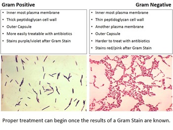
That we're the scientists or pathologists looking Let's talk about how we can actually tell bacteria apart. 7:21 because of their cell wall structure.6:56 has this super-thick peptidoglycan layer,.6:54 But because this Gram positive bacteria.6:33 And because the Gram negative bacteria.6:28 it just stains the whole thing purple,.6:15 before the peptidoglycan layer here.6:10 so next to the plasma membrane space.6:05 And again, think of it peri, next to,.6:02 So, I'm just going to write it over here,.5:51 So above this plasma membrane here.5:49 next to the plasma membrane on both.5:48 And then there's a little bit of space.5:35 which is these lipid areas at the bottom,.5:32 and it's also exactly what it sounds like.5:25 which most people abbreviate as LPS.5:22 also called the lipopolysaccharide layer,.5:19 And then next is this brown layer,.5:15 Then you have an outer membrane, so here.5:11 so I'm not going to write that out again,.4:29 between these rods that you see here.3:56 which are drawn here in little blocks.3:54 with some integral membrane proteins.3:27 compared to Gram positive bacteria,.3:12 It does have the plasma membrane layer,.3:07 So think of this as a zoomed in version.2:59 are really different between Gram positive.2:56 you'll see in a minute that these layers.2:52 So even though right here it says capsule,.2:49 pay attention to just the external layer.2:46 the external layers of the bacteria,.2:43 And again, because the stain is only on.2:38 So this might look familiar to you,.

2:26 the bacteria is called Gram negative.2:14 And if it stains well, it stains purple,.2:06 and this color comes from a special stain.1:59 So now we know about the three main shapes.1:57 and the plural version is spirilla.1:48 And a single rod is called a bacillus,.1:45 or a bunch of them are called cocci.


Current transcript segment: 0:01 - Let's talk about how we can.


 0 kommentar(er)
